In an effort to improve patient safety and save health care dollars, a group of London researchers has turned to a new device for feeding tube insertion.
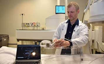
Lawson Health Research Institute clinician researchers teamed up with medical device company CoapTech LLC for the first in the world new method involving 25 patients at the London Health Sciences Centre (LHSC). The new device, called the PUMA-G System, allows the insertion to take place at the patient's bedside. A magnetic balloon is fed through the patient's mouth and then guided down to the stomach using an external magnet. The balloon is then inflated with water. From there, ultrasound is used to guide the doctor as they insert a needle through the stomach and into the balloon. The balloon is then used to catch a wire that is pulled back up to the mouth to remove the balloon. The feeding tube can then be pushed back down over the wire and safely out the stomach.
Traditionally, feeding tubes are inserted using x-ray imaging or endoscopy, a procedure that uses a camera and light to visualize the stomach. While highly effective, this conventional method is costly to the health care system, utilizing busy imaging suites.
"This new method is already showing promise as being safe, effective and efficient. It allows feeding tubes to be inserted at the patient’s bedside and reduces the demand on specialized imaging suites,” said Dr. Derek Cool, associate scientist at Lawson and interventional radiologist at LHSC. “This could be especially important for patients in the intensive care unit. They can benefit from the safety of that environment without being moved.”
Sonny McGlone, 76, of Sarnia was the first patient in the world to undergo the new method of feeding tube insertion. He had lost his ability to eat following radiation therapy for head and neck cancer in October 2018.
“I had seven weeks of radiation, which killed my taste buds. I couldn’t swallow or eat and I was rapidly losing weight,” said McGlone. “I was pleasantly surprised by the feeding tube procedure. While the tube was obviously inconvenient, it was a lifesaver.”
The new device also minimizes the risk of punctured organs by allowing physicians to see between the skin and stomach while the needle is being inserted, according to researchers.
The old method performed in the imaging suite does not show that space and, in rare cases, can lead to liver and colon injuries.
In addition to potentially becoming the new standard for feeding tube insertion, the new device could also be used to provide visualization during other hollow organ procedures.